Understanding Myocardial Perfusion Imaging (MPI) Test: A Comprehensive Overview
Purpose, Procedure, Benefits, and Potential Risks
Introduction: Myocardial Perfusion Imaging (MPI) test is a non-invasive diagnostic procedure used to evaluate blood flow to the heart muscle. It provides valuable insights into the functioning of the heart and aids in the diagnosis of various cardiovascular conditions. In this article, we will explore the purpose, procedure, benefits, and potential risks associated with MPI testing.
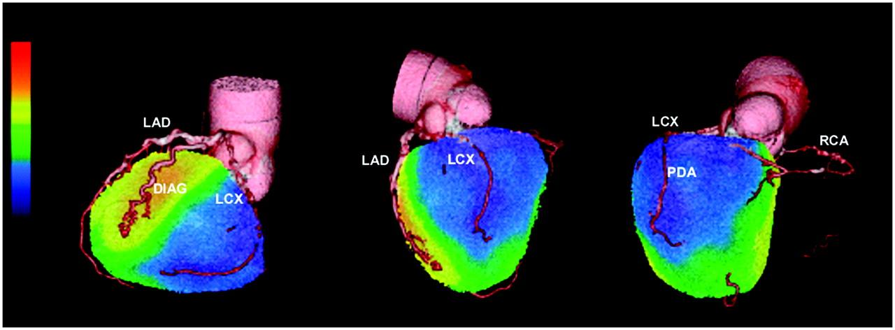
 Purpose of Myocardial Perfusion Imaging (MPI) Test: The MPI test is primarily performed to assess the blood supply to the heart muscle. It helps in detecting coronary artery disease, evaluating the severity of blockages in the coronary arteries, and determining the extent of damage to the heart after a heart attack. The test is also utilized to evaluate the effectiveness of medical or surgical interventions in improving blood flow to the heart.
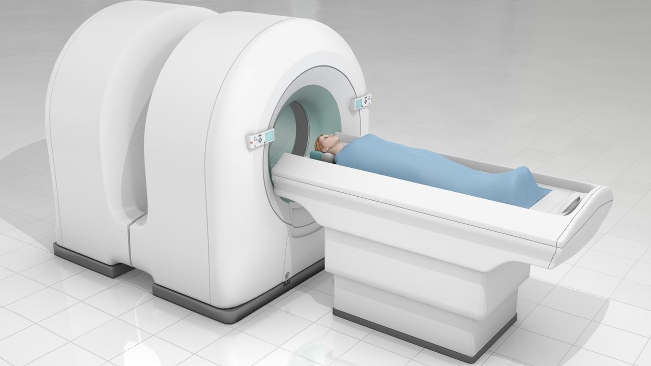
Procedure of Myocardial Perfusion Imaging (MPI) Test: During an MPI test, a small amount of a radioactive substance, usually Technetium-99m or Thallium-201, is injected into the patient's bloodstream. These substances have a short half-life and emit low-level radiation, making them safe for diagnostic purposes. As the radioactive substance circulates through the bloodstream, a specialized camera called a gamma camera or SPECT (Single Photon Emission Computed Tomography) scanner captures images of the heart at rest and during stress.
 To induce stress, the patient may be required to exercise on a treadmill or receive a medication that simulates the effects of exercise on the heart. The stress test helps assess blood flow during periods of increased demand. The images obtained during both the rest and stress phases are then compared to identify any abnormalities in blood flow patterns.
Benefits of Myocardial Perfusion Imaging (MPI) Test: The MPI test offers several benefits for evaluating heart health. It can detect areas of reduced blood flow to the heart, identify the presence and severity of coronary artery disease, and assess the extent of damage caused by a previous heart attack. The test also helps in determining the appropriate treatment strategy, such as medication, angioplasty, or bypass surgery, based on the findings.
Furthermore, MPI testing allows for early detection of cardiac abnormalities, helping to prevent future heart complications and improving patient outcomes. It is a non-invasive procedure that does not involve any surgical intervention or catheterization, making it relatively safe and well-tolerated by most patients.
Potential Risks of Myocardial Perfusion Imaging (MPI) Test: While the MPI test is generally safe, it does involve exposure to a small amount of radiation due to the use of radioactive substances. However, the radiation dose is considered low and poses minimal risk to the patient. The benefits of the test typically outweigh the potential risks.
Conclusion: Myocardial Perfusion Imaging (MPI) test is a valuable diagnostic tool for evaluating blood flow to the heart muscle and detecting various cardiovascular conditions. It provides essential information for diagnosing coronary artery disease, assessing the extent of damage to the heart, and determining the most appropriate treatment options. With its non-invasive nature and minimal risks, MPI testing plays a crucial role in ensuring optimal heart health and improving patient outcomes.
What is a Myocardial Perfusion Imaging (MPI) test?
A MPI test is a diagnostic procedure that uses a radioactive tracer to evaluate blood flow to the heart muscle and identify areas with reduced blood supply.
How is a Myocardial Perfusion Imaging test performed?
During the test, a small amount of radioactive tracer is injected into the bloodstream, and specialized imaging equipment captures images of the heart to assess its blood flow and function.
What are the indications for a Myocardial Perfusion Imaging test?
MPI is commonly used to diagnose coronary artery disease, assess the extent of heart damage after a heart attack, evaluate the effectiveness of treatments, and determine the need for further interventions.
Are there any risks or side effects associated with Myocardial Perfusion Imaging?
The procedure is generally safe, with rare risks of allergic reactions to the tracer. The radiation exposure is minimal and considered safe for most patients.
How long does a Myocardial Perfusion Imaging test take, and when can I expect the results?
The test usually takes about 3-4 hours, including the time for the tracer to circulate in the body. The results are typically available within a few days and will be discussed with your doctor.
We are associated with experienced and highly skilled medical professionals. We use the latest medical technology available in the world and we provide medical services in collaboration with JCI & NABH Certified hospitals only. Our services include various types of treatment and organ restructuring and transplant.
