Understanding Echocardiography with Color Doppler: A Powerful Imaging Technique for Cardiac Assessment
Subheading: Exploring the Applications, Process, and Clinical Benefits of Echocardiography with Color Doppler
Introduction:
Echocardiography with color Doppler is a sophisticated imaging technique widely utilized in modern cardiology. By combining traditional echocardiography with Doppler ultrasound, this non-invasive method enables physicians to visualize the heart's structure, evaluate its function, and assess blood flow patterns in real-time. This article delves into the applications, process, and clinical benefits of echocardiography with color Doppler, highlighting its significance in diagnosing and managing various cardiac conditions.
Understanding Echocardiography with Color Doppler:
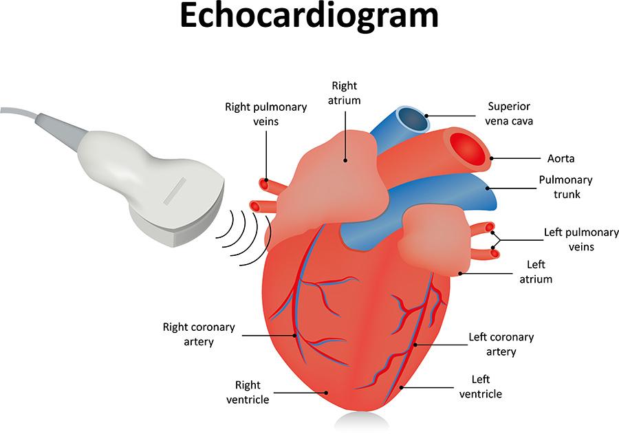
Echocardiography, also known as cardiac ultrasound, employs sound waves to create detailed images of the heart. Color Doppler is an additional feature that allows for the visualization and analysis of blood flow within the heart and blood vessels. By utilizing the Doppler effect, which involves the shift in frequency of sound waves reflected off moving objects, color Doppler can accurately display the direction, velocity, and turbulence of blood flow.

Applications of Echocardiography with Color Doppler:
Assessing Heart Function: Echocardiography with color Doppler provides valuable information about the heart's pumping ability, allowing clinicians to measure parameters such as ejection fraction and cardiac output. It helps in the diagnosis and monitoring of conditions such as heart failure, myocardial infarction, and valvular diseases.
Evaluating Valve Function: Color Doppler enables the assessment of blood flow across heart valves, helping identify valve abnormalities such as stenosis (narrowing) or regurgitation (leakage). This aids in diagnosing conditions like mitral valve prolapse, aortic stenosis, and tricuspid regurgitation.
Detecting Congenital Heart Defects: Echocardiography with color Doppler plays a crucial role in diagnosing congenital heart defects in both children and adults. It provides detailed images of the heart's chambers, septum, and blood vessels, facilitating the identification of conditions such as atrial septal defect, ventricular septal defect, and patent ductus arteriosus.
The Echocardiography with Color Doppler Process:
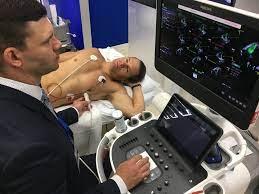
Patient Preparation: The patient lies on an examination table, and small adhesive patches called electrodes are placed on their chest to monitor the heart's electrical activity.
Transducer Placement: A trained sonographer or physician applies a gel to the patient's chest to improve sound wave transmission. They then place a handheld device called a transducer against different areas of the chest, capturing images from different angles.
Image Acquisition: The transducer emits high-frequency sound waves that bounce off the heart structures. The echoes are picked up by the transducer and processed by a computer, generating real-time images of the heart on a monitor.

Color Doppler Analysis: During the examination, the color Doppler feature is activated to visualize blood flow. Directional flow is depicted using different colors, such as red for blood flowing towards the transducer and blue for blood.
We are associated with experienced and highly skilled medical professionals. We use the latest medical technology available in the world and we provide medical services in collaboration with JCI & NABH Certified hospitals only. Our services include various types of treatment and organ restructuring and transplant.
